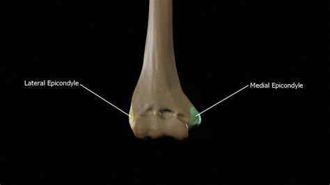
The medial epicondyle of the humerus is a bony projection located on the distal end of the humerus, which is the long bone of the upper arm. This anatomical structure plays a crucial role in the formation of the elbow joint and serves as an attachment point for several muscles and ligaments. The medial epicondyle is situated on the medial (inner) aspect of the humerus, opposite to the lateral epicondyle, and is a key landmark in the diagnosis and treatment of various elbow injuries and conditions.
From an anatomical perspective, the medial epicondyle is a rugged, triangular-shaped prominence that projects medially from the distal end of the humerus. It is the origin of the flexor muscles of the forearm, including the flexor carpi radialis, flexor carpi ulnaris, and flexor digitorum profundus. The medial epicondyle also serves as an attachment point for the ulnar collateral ligament, which provides stability to the elbow joint. The medial epicondyle is separated from the lateral epicondyle by a depression known as the cubital fossa, which contains several important neurovascular structures.
Key Points
- The medial epicondyle of the humerus is a bony projection located on the distal end of the humerus.
- It serves as an attachment point for several muscles and ligaments, including the flexor muscles of the forearm and the ulnar collateral ligament.
- The medial epicondyle plays a crucial role in the formation of the elbow joint and provides stability to the joint.
- It is a key landmark in the diagnosis and treatment of various elbow injuries and conditions, including medial epicondylitis and ulnar collateral ligament injuries.
- The medial epicondyle is separated from the lateral epicondyle by a depression known as the cubital fossa, which contains several important neurovascular structures.
Medial Epicondylitis

Medial epicondylitis, also known as golfer’s elbow, is a common condition characterized by pain and inflammation on the medial (inner) aspect of the elbow. This condition is caused by repetitive strain on the flexor muscles of the forearm, which attach to the medial epicondyle. The repetitive strain leads to microtrauma and inflammation of the tendons, resulting in pain and stiffness in the affected area. Medial epicondylitis is commonly seen in athletes who participate in sports that involve repetitive throwing or gripping motions, such as golf, baseball, and tennis.
Clinical Presentation
The clinical presentation of medial epicondylitis typically includes pain and tenderness on the medial aspect of the elbow, which may radiate down the forearm. Patients may also experience stiffness and limited range of motion in the affected elbow. The pain is often exacerbated by activities that involve gripping, twisting, or flexing the wrist. A thorough physical examination, including palpation and range of motion testing, can help diagnose medial epicondylitis. Imaging studies, such as X-rays or MRI, may be ordered to rule out other conditions, such as fractures or ligament sprains.
| Condition | Symptoms | Treatment |
|---|---|---|
| Medial Epicondylitis | Pain and inflammation on the medial aspect of the elbow | Rest, ice, compression, elevation, physical therapy, and anti-inflammatory medications |
| Ulnar Collateral Ligament Injury | Pain and instability in the medial aspect of the elbow | Rest, ice, compression, elevation, physical therapy, and surgical reconstruction in severe cases |

Ulnar Collateral Ligament Injuries

The ulnar collateral ligament (UCL) is a critical structure that provides stability to the elbow joint. Injuries to the UCL can occur due to repetitive strain or acute trauma, and can result in pain, instability, and limited range of motion in the affected elbow. UCL injuries are common in athletes who participate in sports that involve repetitive throwing or overhead motions, such as baseball, football, and tennis.
Treatment Options
The treatment of UCL injuries depends on the severity of the injury and the patient’s overall health status. Conservative management, including rest, ice, compression, elevation, and physical therapy, may be effective in treating mild to moderate UCL injuries. However, in severe cases, surgical reconstruction of the UCL may be necessary to restore stability and function to the elbow joint. A thorough evaluation of the patient’s symptoms, medical history, and physical examination findings is essential in determining the most effective treatment plan.
In conclusion, the medial epicondyle of the humerus is a complex anatomical structure that plays a critical role in the formation and function of the elbow joint. Medial epicondylitis and UCL injuries are common conditions that can cause significant pain and disability, and require a thorough understanding of the anatomy and biomechanics of the elbow joint to diagnose and treat effectively. A combination of conservative management and surgical intervention, when necessary, can help alleviate symptoms and promote healing in patients with these conditions.
What is the medial epicondyle of the humerus?
+The medial epicondyle of the humerus is a bony projection located on the distal end of the humerus, which serves as an attachment point for several muscles and ligaments, including the flexor muscles of the forearm and the ulnar collateral ligament.
What is medial epicondylitis?
+Medial epicondylitis, also known as golfer’s elbow, is a common condition characterized by pain and inflammation on the medial (inner) aspect of the elbow, caused by repetitive strain on the flexor muscles of the forearm.
What are the symptoms of UCL injuries?
+The symptoms of UCL injuries include pain, instability, and limited range of motion in the affected elbow, and can be caused by repetitive strain or acute trauma.