The animal cell is a complex and highly organized structure that is composed of various organelles, each with its own unique function. Understanding the components of an animal cell is crucial for grasping the fundamental principles of biology and how living organisms function. In this article, we will delve into the details of an animal cell labeled diagram, exploring the different parts of the cell and their roles in maintaining cellular homeostasis.
Introduction to Animal Cells

Animal cells are eukaryotic cells, meaning their DNA is enclosed within a nucleus. They are found in a wide range of organisms, from simple sponges to complex mammals. Unlike prokaryotic cells, which lack a nucleus and other membrane-bound organelles, animal cells are characterized by their intricate internal structure. This complexity allows animal cells to perform a variety of functions necessary for the survival of the organism.
Key Points
- The animal cell is a type of eukaryotic cell that contains a nucleus and various organelles.
- Each organelle within the animal cell has a specific function that contributes to the overall health and operation of the cell.
- Understanding the structure and function of animal cells is essential for understanding biological processes and addressing health issues.
- Animal cells can vary in shape, size, and the number of organelles, depending on their specific functions within the organism.
- The ability of animal cells to differentiate and specialize is a key factor in the development and complexity of multicellular organisms.
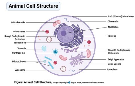
Components of an Animal Cell

An animal cell labeled diagram includes several key components, each playing a vital role in cellular function. These components include:
1. Plasma Membrane
The plasma membrane, also known as the cell membrane, is the outermost layer of the cell. It is a semi-permeable membrane that regulates the movement of substances in and out of the cell. The plasma membrane is composed of a phospholipid bilayer with embedded proteins that perform various functions, including transport, signaling, and cell-cell recognition.
2. Cytoplasm
Cytoplasm is the jelly-like substance within the cell, enclosing the organelles. It is composed of water, salts, sugars, and various organelles. The cytoplasm is the site where many metabolic reactions occur and serves as a medium for the exchange of materials between different parts of the cell.
3. Nucleus
The nucleus is the control center of the cell, containing most of the cell’s genetic material in the form of DNA. It is surrounded by a double membrane called the nuclear envelope, which has nuclear pores that allow for the passage of materials between the nucleus and the cytoplasm. The nucleus plays a crucial role in cell growth, metabolism, and reproduction by controlling gene expression.
4. Mitochondria
Mitochondria are often referred to as the “powerhouses” of the cell because they generate most of the cell’s supply of adenosine triphosphate (ATP), which is used as a source of chemical energy. They are unique in having their own DNA and can reproduce independently of the cell cycle.
5. Endoplasmic Reticulum (ER)
The endoplasmic reticulum is a network of membranous tubules within the cytoplasm of the cell. It comes in two forms: rough ER, which is studded with ribosomes and involved in protein synthesis, and smooth ER, which is involved in lipid synthesis and detoxification.
6. Lysosomes
Lysosomes are membrane-bound vesicles that contain digestive enzymes. They are responsible for cellular digestion and recycling of cellular waste and foreign substances that enter the cell.
7. Golgi Apparatus
The Golgi apparatus, or Golgi complex, is a complex organelle found in most eukaryotic cells. It is responsible for modifying, sorting, and packaging proteins and lipids for storage or transport out of the cell.
8. Cytoskeleton
The cytoskeleton is a complex network of filaments that provides structural support, shape, and mechanical stability to cells. It is composed of three main components: microtubules, microfilaments, and intermediate filaments, each with distinct functions in cell division, movement, and intracellular transport.
| Organelle | Function |
|---|---|
| Plasma Membrane | Regulates the movement of substances in and out of the cell |
| Cytoplasm | Serves as a medium for metabolic reactions and material exchange |
| Nucleus | Contains genetic material and controls gene expression |
| Mitochondria | Generates ATP for the cell |
| Endoplasmic Reticulum | Involved in protein and lipid synthesis |
| Lysosomes | Responsible for cellular digestion and recycling |
| Golgi Apparatus | Modifies, sorts, and packages proteins and lipids |
| Cytoskeleton | Provides structural support and mechanical stability |

Conclusion
In conclusion, an animal cell labeled diagram reveals the intricate and highly specialized structure of eukaryotic cells. Each component, from the plasma membrane to the cytoskeleton, plays a vital role in the cell’s operation, contributing to the organism’s overall health and function. By studying the animal cell and its various organelles, we can gain a deeper understanding of biological processes and develop new insights into the treatment of diseases.
What is the primary function of the nucleus in an animal cell?
+The primary function of the nucleus is to contain the cell’s genetic material in the form of DNA and to control gene expression, thereby regulating cell growth, metabolism, and reproduction.
What is the role of mitochondria in animal cells?
+Mitochondria are responsible for generating most of the cell’s supply of adenosine triphosphate (ATP), which is used as a source of chemical energy for various cellular processes.
What is the difference between rough and smooth endoplasmic reticulum?
+Rough endoplasmic reticulum is studded with ribosomes and is involved in protein synthesis, whereas smooth endoplasmic reticulum is involved in lipid synthesis and detoxification.